Tutorial: Evolution of Microwave and Millimeter Wave Imaging for NDE Applications & Diagnosis of Human Skin Lesions (Cancer and Burns) Using High-Frequency Techniques

You are here

Evolution of Microwave and Millimeter Wave Imaging for NDE Applications & Diagnosis of Human Skin Lesions (Cancer and Burns) Using High-Frequency Techniques

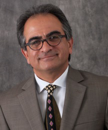
Reza Zoughi

Applied Microwave Nondestructive Testign Laboratory (amntl), Electrical and Computer Engineering Department, Missouri University of Science and Technology (S & T), Rolla, MO 64509


ABSTRACT
Millimeter wave signals span the frequency range of 30 GHz to 300 GHz, corresponding to a wavelength range of 10 mm to 1 mm. Signals at these frequencies are non-ionizing and easily penetrate inside dielectric materials and composites and interact with their inner structures. The relatively small wavelengths and wide bandwidths associated with these signals enable production of high spatial-resolution images of materials and structures. Incorporating imaging techniques based on robust back-propagation algorithms coupled with more advanced and unique millimeter wave imaging systems have brought upon a flurry of activities in this area and in particular for nondestructive evaluation (NDE) applications. Ultimately, imaging techniques must produce high-resolution (in 3D) holographical images, become real-time, and be implemented using portable systems.  To this end, recently the design and demonstration of a 6” by 6” one-shot, rapid and portable imaging system (Microwave Camera), consisting of 576 resonant slot elements, was completed.  Subsequently, successful efforts have been expended to design and implement several different variations of this imaging system, notably one that is capable of producing real-time 3D images.

High-frequency signals in the microwave and millimeter wave regions are also effective candidates for evaluating human skin lesions caused by burns and cancers. According to the American Cancer Society (ACS) “Cancer of the skin is by far the most common of all cancers. Melanoma accounts for less than 2% of skin cancers cases but causes a large majority of skin cancer deaths”.  The ACS estimates that in 2014 in the United States about 76,100 new cases of melanoma will have been diagnosed and approximately 9,710 people are expected to die from melanoma. If diagnosed in their early stages, 95% skin cancers are curable. Visual inspection using size, shape, color, border irregularities, ulceration, tendency to bleed and whether the lesion is raised, hard or tender are common approaches to diagnosis. Visual inspection is subjective and susceptible to human error. Malignant skin tumors have different biological properties than the surrounding healthy skin, which enables distinction between these two types of skin using a proper inspection technique. A noninvasive method producing reliable and real-time information about a suspected skin malignancy, that enables dermatologists to obtain a real-time diagnosis of the likelihood of a lesion being cancerous, would be of great clinical and diagnostic value.

Burn injury represents a wide range of tissue damage. The classification and treatment of thermal injuries are determined based on the depth of invasion into the underlying tissue. The postoperative management of skin and skin-substitute grafts is complicated by the need to stabilize the grafts with dressings, which introduces some limitations for readily removing it to monitor the grafted wound for correctible problems.  When it comes to burned skin, comprehensive diagnosis refers to detection as well as evaluation of critical parameters, the most critical of which is the depth of invasion. A diagnostic tool allowing for real-time qualitative and quantitative evaluation of a burn through desiccated skin or optically-opaque dressings represents a significant addition to the medical toolbox used by physicians and first responders caring for burned patients.

As it relates to evaluation of human skin, the interaction of millimeter wave signals is dependent upon the biophysical (i.e., dielectric and thickness) properties of skin, as well as electromagnetic parameters such as the frequency of operation and specific characteristics of the probe used. There are several technical and practical beneficial features that make high-frequency evaluation of human skin quite attractive as a potential medical diagnostics tool. The possibility of real-time imaging of human skin at millimeter wave frequencies, has the real potential to lead to a new paradigm shift in the way human skin is diagnosed for burns and cancers.

In this tutorial, a historical and technical review of high-frequency inspection techniques, used for evaluating skin cancer and burned skin, will be presented. Issues related to technical advances in developing real-time imaging systems as well as the potential future possibilities in this realm will be discussed.

SPEAKER BIOGRAPHY
R. Zoughi received his B.S.E.E, M.S.E.E, and Ph.D. degrees in electrical engineering (radar remote sensing, radar systems, and microwaves) from the University of Kansas where from 1981 until 1987 he was at the Radar Systems and Remote Sensing Laboratory (RSL). Subsequently, in 1987 he joined the Department of Electrical and Computer Engineering at Colorado State University (CSU), where he established the Applied Microwave Nondestructive Testing Laboratory (amntl). He held the position of Business Challenge Endowed Professor of Electrical and Computer Engineering from 1995 to 1997 while at CSU.  In 2001 he joined the Department of Electrical and Computer Engineering at Missouri University of Science and Technology (S&T), formerly University of Missouri-Rolla (UMR), as the Schlumberger Distinguished Professor.  His current areas of research include developing new nondestructive techniques for microwave and millimeter wave testing and evaluation of materials (NDT&E), developing new electromagnetic probes and sensors to measure characteristic properties of material at microwave frequencies, developing embedded modulated scattering techniques for NDT&E purposes and real-time high resolution imaging system development. He is the author of a book entitled “Microwave Nondestructive Testing and Evaluation Principles”, and the co-author of a chapter on Microwave Techniques in an undergraduate introductory textbook entitled “Nondestructive Evaluation: Theory, Techniques, and Applications”.  He has been the recipient of numerous teaching awards both at CSU and Missouri S&T.  He is the co-author of over 560 journal papers, conference proceedings and presentations and technical reports. He has fifteen patents to his credit all in the field of microwave nondestructive testing and evaluation. He was the recipient of the 2007 IEEE Instrumentation and Measurement Society Distinguished Service Award, the 2009 American Society for Nondestructive Testing (ASNT) Research Award for Sustained Excellence, and the 2011 IEEE Joseph F. Keithley Award in Instrumentation & Measurement.  In 2013 he and his co-authors received the H. A. Wheeler Prize Paper Award of the IEEE Antennas and Propagation Society. He is a Fellow of the Institute of Electrical and Electronics Engineers (IEEE), a Fellow of American Society for Nondestructive Testing (ASNT), and served as the Editor-in-Chief of the IEEE Transactions on Instrumentation and Measurement from 2007-2011.  He also served as the President of the IEEE Instrumentation and Measurement Society (2014-2015).